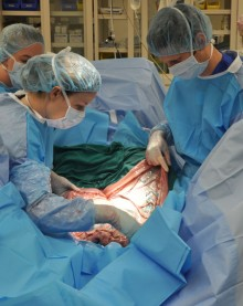
CALEC surgery, a groundbreaking procedure developed at Mass Eye and Ear, is redefining the landscape of ocular repair by leveraging stem cell therapy to address serious corneal damage. This innovative approach involves extracting limbal epithelial cells from a healthy eye and cultivating them into grafts that can restore the corneal surface in patients suffering from previously untreatable injuries. In a recent clinical trial, CALEC surgery demonstrated remarkable efficacy, with over 90% of participants experiencing significant improvements in vision and quality of life. The implications of this stem cell treatment extend beyond immediate restoration, suggesting potential avenues for comprehensive ocular surface repair amid a pressing need for novel therapies. As research continues to evolve, CALEC surgery stands as a beacon of hope for those facing the challenges of corneal damage, promoting the advancement of transformative solutions in eye care.
The cultivated autologous limbal epithelial cell (CALEC) approach represents a significant innovation in corneal restoration methods, paving the way for new possibilities in ocular surface rehabilitation. This procedure not only focuses on the extraction and expansion of vital limbal stem cells but also emphasizes the integration of advanced manufacturing techniques to create viable grafts for transplantation. By utilizing stem cell-derived solutions, researchers aim to address the challenges previously faced by patients with severe corneal damage from various injuries or conditions. This methodology, part of a comprehensive strategy for ocular health, is fundamentally changing how we understand and treat corneal ailments, illustrating a robust paradigm shift in regenerative medicine within ophthalmology. With its promising results, CALEC surgery highlights the synergy between scientific innovation and compassionate care in addressing complex eye diseases.
The Breakthrough of CALEC Surgery in Eye Care
CALEC surgery, or Cultivated Autologous Limbal Epithelial Cells surgery, represents a significant advancement in the field of ophthalmology, specifically for treating corneal damage. This innovative technique involves extracting stem cells from a healthy part of a patient’s eye and cultivating them to produce a graft that can be transplanted onto the damaged eye. The procedure addresses the pressing need for effective treatments for severe corneal injuries, such as those caused by chemical burns or other traumas, which traditionally have limited treatment options.
At Mass Eye and Ear, CALEC surgery has shown promising results. In a clinical trial, researchers reported a success rate exceeding 90% in restoring the cornea’s surface, which has led to improved vision and quality of life for those affected. As noted by Ula Jurkunas, the principal investigator of the trial, the outcomes go beyond mere restoration; they denote a substantial advancement in ocular surface repair, setting a new standard for treatments that were once considered futile.
How CALEC Surgery Works: A Step-by-Step Process
The CALEC surgical process begins with a biopsy of limbal epithelial cells taken from a healthy eye. These vital stem cells are crucial for maintaining the integrity of the corneal surface. The harvested cells are then cultured and expanded in a lab environment to create a cellular tissue graft. This innovative manufacturing method takes approximately two to three weeks, during which the cells multiply and form the necessary graft tissue.
Once the graft is ready, it is surgically transplanted onto the damaged cornea, effectively restoring its surface. The meticulous approach to creating and applying this graft exemplifies the marriage between advanced biotechnology and traditional surgical techniques. The results are encouraging, with over half of the trial participants experiencing complete restoration of their cornea within three months post-surgery.
Throughout the clinical trials at Mass Eye and Ear, the safety and feasibility of CALEC surgery have been closely monitored. The absence of significant adverse events highlights the procedure’s promise, paving the way for broader applications and additional research into stem cell therapies.
Understanding the Role of Stem Cells in Corneal Repair
Stem cell therapy has emerged as a revolutionary approach in treating various eye conditions, particularly those involving corneal damage. The eye’s limbus contains a reservoir of limbal epithelial stem cells that play a crucial role in maintaining the cornea’s health and structure. When these cells are depleted due to injury or disease, the corneal surface becomes unstable, leading to vision impairment and persistent pain.
The utilization of stem cells in CALEC surgery allows for the regeneration of the corneal surface, providing hope for patients who previously faced a bleak prognosis. This groundbreaking technique harnesses the body’s natural capacity for healing, making it a standout innovation in ocular care. By focusing on restoring the limbal epithelial cells, CALEC not only repairs the eye but also addresses associated complications from impaired ocular surfaces.
The Importance of Clinical Trials in Advancing Eye Treatments
Clinical trials are pivotal in validating new treatments such as CALEC surgery, ensuring they are both safe and effective before they can be widely adopted in clinical practice. The initial clinical trial led by Ula Jurkunas at Mass Eye and Ear involved rigorous evaluation of the therapy over an 18-month period, demonstrating significant benefits for patients suffering from severe corneal damage. This step signifies critical progress toward establishing protocols for future treatments that prioritize evidence-based care.
Further, the successful outcomes and data derived from these trials can guide further research and clinical developments. The hope is that, as CALEC surgery gains attention and support, it may inspire larger studies across multiple centers—a model that has been essential in bringing advanced therapies to fruition, enhancing patient care in ophthalmology.
Potential Future Directions for CALEC Research
Research on CALEC surgery reflects an evolving understanding of stem cell therapies in ocular treatments. Future studies aim to expand on the initial work by involving larger patient cohorts and exploring the potential of allogeneic stem cell sources, such as cadaveric donor eyes. This could significantly impact how corneal damage is treated, especially for patients with injuries affecting both eyes.
Continued collaboration among research institutions like Mass Eye and Ear, Dana-Farber, and Boston Children’s Hospital will be crucial in refining the techniques and processes surrounding CALEC surgery. By building on existing studies, the goal is to not only further validate the effectiveness of this treatment but also to seek FDA approval, making it accessible to a broader patient population.
Impact of CALEC Surgery on Quality of Life
For many patients with corneal damage, the consequences extend beyond visual impairments to include chronic pain and reduced quality of life. The results from the CALEC surgery trials indicate that restoring the corneal surface significantly alleviates these issues, leading to improved visual acuity and overall life satisfaction. Success stories from trial participants underline the transformative power of this innovative treatment.
The psychological and emotional benefits of restoring vision through CALEC can be profound. Patients often report increased social interactions, improved occupational performance, and heightened personal autonomy after successful treatment. By focusing on both the physiological and psychosocial aspects of eye care, CALEC surgery stands as a comprehensive solution for individuals suffering from debilitating corneal injuries.
Collaborative Efforts in Advancing Ocular Therapy
The development of CALEC surgery exemplifies the power of collaborative research in advancing medical technology. Through the partnership of Mass Eye and Ear with institutions such as Dana-Farber and Boston Children’s Hospital, researchers have been able to push the boundaries of what is possible in treating corneal damage. This collaborative environment fosters innovation and ensures rigorous scientific standards are met.
By pooling resources, expertise, and technological capabilities, these institutions have made significant strides in creating a robust framework for testing and validating new treatments. Future efforts should continue to emphasize collaboration, as this approach will be pivotal in identifying effective therapies that can be reliably rolled out for patient care.
Challenges and Limitations of CALEC Surgery
While CALEC surgery demonstrates great promise, it is not without its challenges. One significant limitation is the requirement of having a healthy eye from which to extract stem cells, restricting eligibility solely to patients with unilateral corneal damage. This condition limits the number of individuals who can benefit from this innovative therapy.
Additionally, as with any surgical procedure, there are inherent risks that must be considered. Although the trial noted a high safety profile, ongoing monitoring and evaluation are essential as treatments progress from clinical trials to everyday clinical settings. Addressing these challenges will be crucial for the advancement and acceptance of CALEC surgery as a standard practice in ocular care.
The Role of the National Eye Institute in CALEC Research
The National Eye Institute (NEI) has played a vital role in funding and supporting the clinical trials for CALEC surgery. By investing in such innovative research, the NEI helps pave the way for breakthroughs that can change the landscape of ocular treatments. Their commitment to understanding and improving eye health is integral to developing new therapies that provide hope to patients with previously untreatable conditions.
Through partnerships with leading research centers, the NEI is not only enhancing the scientific community’s understanding of corneal repair but also ensuring that promising therapies undergo rigorous testing to meet the highest standards. This ongoing support is essential in transitioning innovative treatments from the research phase to clinical application, ultimately benefiting patients across the United States.
Frequently Asked Questions
What is CALEC surgery and how does it relate to stem cell therapy?
CALEC surgery, or Cultivated Autologous Limbal Epithelial Cell surgery, is an innovative procedure developed at Mass Eye and Ear that utilizes stem cell therapy to treat serious corneal injuries. This surgery involves transplanting limbal epithelial cells, which are extracted from a healthy eye, to repair the ocular surface of a damaged eye. By restoring the cornea’s surface, CALEC surgery has shown over 90% effectiveness in treating patients with previously untreatable corneal damage.
How does CALEC surgery help in the treatment of corneal damage?
CALEC surgery helps treat corneal damage by regenerating the limbal epithelial cells that are essential for eye health. During the procedure, these cells are harvested from a healthy part of the eye and cultivated into a graft, which is then transplanted into the affected eye. This innovative approach has been shown to restore the corneal surface effectively, significantly improving the quality of life for patients suffering from severe ocular surface damage.
What are limbal epithelial cells and why are they important for ocular surface repair in CALEC surgery?
Limbal epithelial cells are specialized stem cells located at the limbus, the border of the cornea, that play a crucial role in maintaining the smooth surface of the eye. They are vital for ocular surface repair because injuries to the cornea can deplete these cells, leading to persistent pain and vision issues. CALEC surgery successfully utilizes these cells to regenerate the corneal surface, making it an essential treatment for patients with severe corneal injuries.
Can anyone with corneal damage undergo CALEC surgery?
Currently, CALEC surgery is suited for patients who have healthy limbal epithelial cells in one eye. The procedure requires harvesting cells from the unaffected eye via biopsy. Patients with damage to both eyes may not be eligible at this stage; however, researchers are exploring future methods, such as using cadaveric donor eyes, to broaden the eligibility for CALEC surgery.
What are the expected outcomes and success rates for patients undergoing CALEC surgery?
In clinical trials, CALEC surgery demonstrated a success rate of approximately 90% in restoring corneal surfaces. Specifically, half of the participants had complete restoration of the cornea at three months, with success rates rising to 79% and 77% at 12 and 18-month follow-ups, respectively. The surgery not only aims for visual restoration but also significantly enhances the overall quality of life for patients.
Is CALEC surgery currently available for patients outside of clinical trials?
As of now, CALEC surgery remains experimental and is not widely available at Mass Eye and Ear or other U.S. hospitals. Ongoing studies are needed to further assess its safety and effectiveness before it can be submitted for federal approval. Patients interested in this innovative therapy should consult their ophthalmologist for the latest information.
What makes CALEC surgery a unique approach in the treatment of eye injuries?
CALEC surgery stands out as a unique approach because it harnesses the power of stem cell therapy to address corneal damage that was previously deemed untreatable. By cultivating limbal epithelial cells from a healthy eye and transplanting them into a damaged one, this innovative treatment is at the forefront of regenerative medicine for eye health, promising new hope for restoration.
| Key Points |
|---|
| Ula Jurkunas performs the first CALEC surgery at Mass Eye and Ear. |
| A clinical trial successfully restored corneal surfaces in 14 patients over 18 months. |
| Cultivated autologous limbal epithelial cells (CALEC) helps repair serious corneal injuries. |
| The method involves extracting stem cells from a healthy eye, expanding them, and transplanting them. |
| The procedure is over 90% effective, significantly improving patients’ quality of life. |
| The FDA approved the clinical trial, marking the first human study of this therapy in the U.S. |
| Future research aims to treat patients with damage in both eyes using cadaveric stem cells. |
| High safety profile with limited minor adverse events reported. |
| Further studies are necessary before nationwide availability of CALEC therapy. |
Summary
CALEC surgery offers a groundbreaking treatment for individuals with serious corneal damage, highlighting a promising advancement in ophthalmology. The clinical trial led by Ula Jurkunas demonstrates that this innovative stem cell therapy is not only safe but also remarkably effective, boasting a 90% success rate in restoring corneal surfaces. As researchers work toward expanding CALEC’s applications, the future looks bright for those suffering from previously untreatable eye conditions.
